Animal Cell Division Mitosis Model
Product Code : EL-ZM-10609
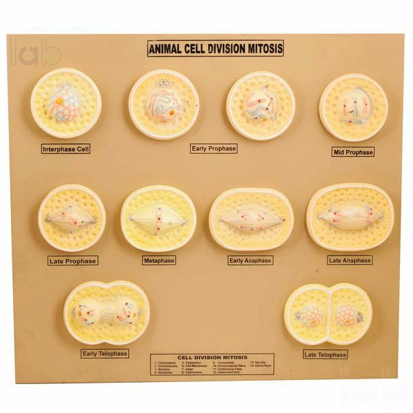
Quick answer: The Animal Cell Division Mitosis Model is a set of 10 models that shows mitotic cell division in an animal cell and its various stages. It is used to teach how a cell copies and separates its chromosomes so that both daughter cells receive a full set. The chromosomes are painted for easy identification. Lab Exports manufactures it in Delhi for schools, colleges and universities.
Specifications
| Parameter | Detail |
|---|---|
| Set | 10 models |
| Coverage | Process of mitotic cell division and its various stages |
| Chromosomes | Painted for easy identification |
| Identification | Numbered with English key card |
How the model set is used in teaching
Mitosis is the division of a body cell into two daughter cells with the same chromosome number as the parent. Textbooks describe prophase, when the chromosomes condense and the nuclear envelope breaks down; metaphase, when they line up at the equator of the spindle; anaphase, when the sister chromatids move to opposite poles; and telophase, when new nuclei form, followed by cytokinesis, which in an animal cell is a furrow that pinches the cell in two.
A set of ten separate models lets the teacher lay the process out in order on the bench, so students see the change from one stage to the next instead of a single diagram. Because the chromosomes are painted, their position and behaviour can be followed across the whole sequence.
- Arrange the ten models in order and use the English key card to name the numbered features.
- Ask students to identify each stage from the position of the painted chromosomes.
- Compare the models with a prepared mitosis slide under the microscope.
- Contrast the sequence with meiosis to show why mitosis keeps the chromosome number constant.
Key features
- Set of 10 models covering the stages of mitotic cell division
- Chromosomes painted for easy identification at every stage
- Numbered features identified on an English key card
- Separate models can be arranged in sequence for step-by-step teaching
- Suitable for cytology, genetics and cell biology lessons
Documentation and supply
An English key card for the numbered features is supplied with the set. Most Lab Exports products carry a one-year manufacturer warranty.
Lab Exports manufactures the mitosis model set at its works in Laxmi Nagar, Delhi, and supplies it to schools, colleges and universities in India and 60+ countries, handling the export documents for each shipment. More about the company is on the About Lab Exports page.
Related Zoological Models products
- RNA Model (EL-ZM-10608)
- DNA Model Kit (EL-ZM-10607)
- Locust Anatomy Model (EL-ZM-10606)
- Euglena Model (EL-ZM-10605)
- Paramecium Model (EL-ZM-10604)
- Head of the Pork Tape Worm Model (EL-ZM-10603)
The full range is in the Zoological Models section of the Biology Lab range.
Frequently asked questions
How many models are in the mitosis model set?
The set contains 10 models showing the process of mitotic cell division and its various stages.
How are the chromosomes shown on the mitosis models?
The chromosomes are painted so that they are easy to identify and to follow from one stage to the next.
What is the price of the mitosis model set?
Prices are not published on the website. Add the mitosis model set to the Enquiry Cart with the Add To Cart button on this page, or email [email protected] with the house code EL-ZM-10609 and the quantity, to receive a quotation.
Does Lab Exports manufacture the mitosis model set?
Yes. Lab Exports manufactures the mitosis model set at its works at 11/315, Lalita Park, Laxmi Nagar, Delhi 110092, under house code EL-ZM-10609.
Request a quote
Email [email protected] with the house code EL-ZM-10609 and the quantity, or add the mitosis model set to the Enquiry Cart with the Add To Cart button on this page. Tender buyers can also see the Tenders and OEM page.
Model.jpg)