Hydra Model
Product Code : EL-ZM-10601
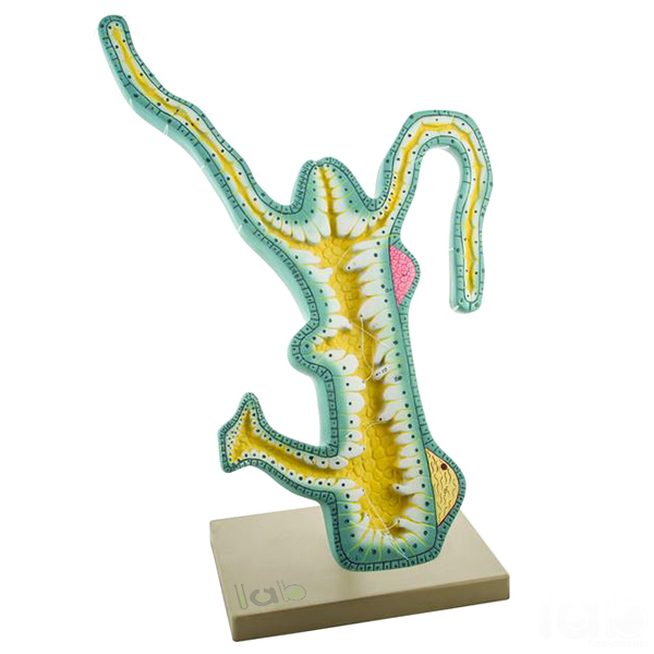
Quick answer: The Hydra Model is a colourfully illustrated model on a stand showing the external and internal structure of hydra, including the bud, tentacles, hypostome, mouth, nematocysts, epidermis, mesoglea, gastrodermis, gastrovascular cavity, testis, ovary and foot. It is used to teach cnidarian structure and reproduction. Lab Exports manufactures it in Delhi for schools, colleges and universities.
Specifications
| Parameter | Details |
|---|---|
| Structures shown | Bud, gastrovascular cavity, testis, tentacle, foot, ovary, hypostome, mesoglea, mouth, nematocysts, epidermis, gastrodermis, flagellated cell |
| Presentation | Colourfully illustrated |
| Mounting | On stand |
| Identification | Numbered, with English key card |
How the model is used in teaching
Hydra is a tiny freshwater animal of the phylum Cnidaria, only a few millimetres long, so its structure is hard to see without a microscope. Its tube-shaped body has two cell layers, the outer epidermis and the inner gastrodermis, with a jelly-like mesoglea between them. At the top, a raised hypostome carries the mouth, surrounded by tentacles armed with stinging nematocysts that capture small prey.
Hydra reproduces in two ways, and the model shows both: asexually by budding, where a small new hydra grows from the side of the body, and sexually through testes and ovaries on the body wall. Enlarged and coloured, the model lets a whole class follow these structures together.
- Identify the foot, body column, hypostome and tentacles.
- Locate the nematocysts on the tentacles and explain how they capture prey.
- Compare the epidermis, mesoglea and gastrodermis of the body wall.
- Point out the bud, testis and ovary and compare asexual with sexual reproduction.
Key features
- External and internal structure of hydra in one model
- Bud, testis and ovary for teaching both types of reproduction
- Epidermis, mesoglea and gastrodermis shown
- Nematocysts and flagellated cell detail
- Colourfully illustrated, on a stand, with an English key card
Documentation and supply
The numbered structures are explained on an English key card supplied with the model. Most Lab Exports products carry a one-year manufacturer warranty.
Lab Exports manufactures the hydra model at its works in Laxmi Nagar, Delhi, and supplies it to schools, colleges and universities in India and 60+ countries, handling the export documents for each shipment. More about the company is on the About Lab Exports page.
Related Zoological Models products
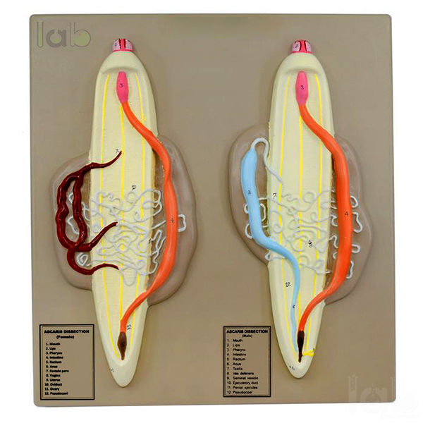
- Ascaris Male and Female Model (EL-ZM-10600)
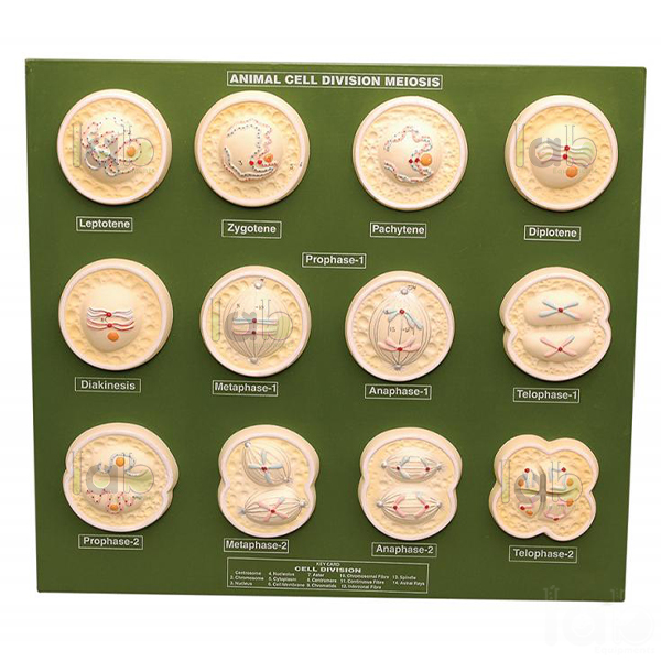
- Animal Cell Division Meiosis Model (EL-ZM-10602)
- Rat Dissection Female Model (EL-ZM-10599)
- Head of the Pork Tape Worm Model (EL-ZM-10603)
- Cockroach Dissection Male Model (EL-ZM-10598)
- Paramecium Model (EL-ZM-10604)
The full range is in the Zoological Models section of the Biology Lab range.
Frequently asked questions
What structures does the hydra model show?
The bud, tentacles, hypostome, mouth, nematocysts, epidermis, mesoglea, gastrodermis, flagellated cell, gastrovascular cavity, testis, ovary and foot.
Does the hydra model show reproduction?
Yes. It shows a bud for asexual reproduction and the testis and ovary for sexual reproduction.
What is the price of the hydra model?
Prices are not published on the website. Add the hydra model to the Enquiry Cart with the Add To Cart button on this page, or email [email protected] with the house code EL-ZM-10601 and the quantity, to receive a quotation.
Does Lab Exports manufacture the hydra model?
Yes. Lab Exports manufactures the hydra model at its works at 11/315, Lalita Park, Laxmi Nagar, Delhi 110092, under house code EL-ZM-10601.
Request a quote
Email [email protected] with the house code EL-ZM-10601 and the quantity, or add the hydra model to the Enquiry Cart with the Add To Cart button on this page. Tender buyers can also see the Tenders and OEM page.